
When we think of medical imaging, the first thing that comes to mind is often the X-ray. And while X-rays are fantastic for looking at broken bones or air-filled lungs, they have a blind spot, which is soft tissue.
On an X-ray, the liver, spleen, kidneys, and bladder all look like similar shades of grey. To tell them apart—and to see inside them—we need a different tool.
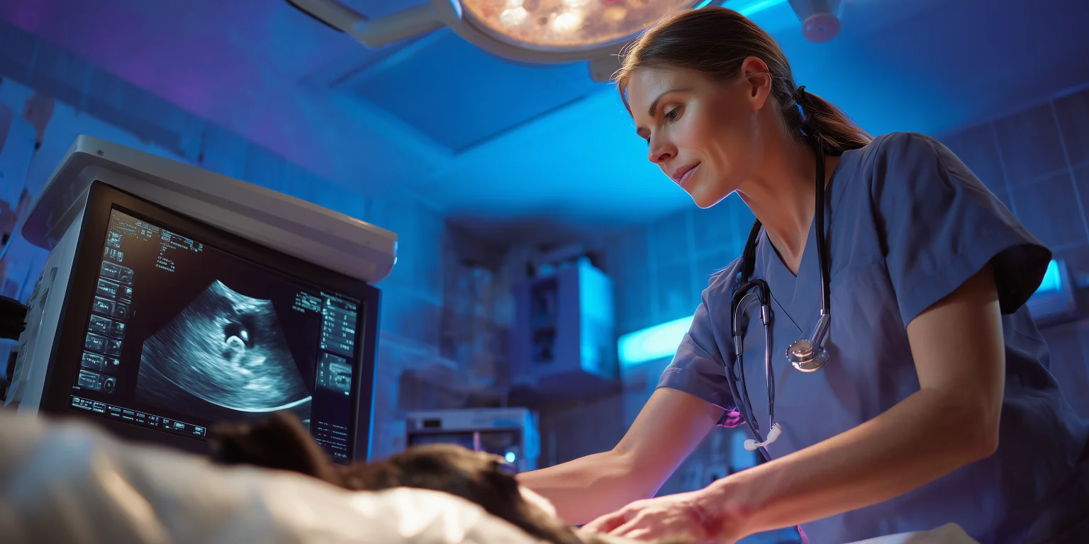
At North Hill Veterinary Clinic, we utilize advanced Veterinary Ultrasound to look inside your pet’s organs in real-time. It is the same technology used to scan babies in the womb, adapted for our four-legged patients.
What is Veterinary Ultrasound?
Unlike X-rays, which use radiation, Ultrasound uses high-frequency sound waves.
A handheld probe (transducer) emits sound waves into the body. These waves bounce off the organs and return to the probe like an echo. The computer translates these echoes into a moving, detailed image on a screen.
Because it is a “movie” rather than a “photo,” it allows us to see:
Architecture: We can see the texture of the liver or kidney (looking for tumors or cysts).
Movement: We can watch the heart beating or the intestines contracting (peristalsis).
Flow: Using “Doppler,” we can see blood flowing through vessels to check for clots or shunts.
When Do We Use an Ultrasound at Armidale Vet?
Ultrasound answers questions that blood tests and X-rays cannot.
1. The “Big Belly” (Abdominal Scan)
If your pet has a swollen abdomen or abnormal liver enzymes, an ultrasound allows us to tour the entire abdomen. We check the:
Liver & Spleen: For masses or tumors.
Kidneys: For size, shape, and signs of chronic failure.
Bladder: For stones, tumors, or wall thickening (cystitis).
Adrenals: For Cushing’s disease tumors.
2. Pregnancy Diagnosis
Just like in humans, this is the most joyful use of the machine! We can confirm pregnancy from around Day 28–30 after mating. We can see the fetal heartbeats to confirm the puppies or kittens are alive and viable.
Note: For an exact count of how many puppies, we still recommend an X-ray at Day 55.
3. Heart Disease (Echocardiogram)
This is a specialized ultrasound of the heart. It allows us to measure the thickness of the heart walls and watch the valves opening and closing. It is essential for diagnosing heart murmurs and Dilated Cardiomyopathy (DCM).
4. Guided Biopsies
If we find a mass on the liver or spleen, we don’t always need open surgery to identify it. Using the ultrasound screen as a guide, we can guide a needle precisely into the mass to take a sample (Tru-Cut Biopsy) without hitting any major blood vessels.
What to Expect During Your Visit
The procedure is very different from an X-ray. It requires patience, darkness, and a little bit of grooming.
1. The “Reverse Mohawk” (Shaving)
This is the part owners are often most surprised by! Ultrasound waves cannot travel through air or fur. To get a clear image, the probe must make perfect contact with the skin.
We must shave a square patch on your pet’s belly (or chest for heart scans).
Don’t worry—it grows back!
2. The Gel
We apply plenty of water-soluble acoustic gel to the skin. It feels a bit cold and wet, but it is necessary to conduct the sound waves.
3. The Procedure
Your pet usually lies on a soft, V-shaped cushion on their back. The room lights are dimmed so the vet can see the screen clearly.
Is it painful? Not at all. In fact, many dogs enjoy the tummy rub!
Sedation: Most pets are happy to lie still with some gentle reassurance. However, if your pet is very anxious or painful, we may give a light sedative to help them relax.