We have all been there: you are cuddling your dog or cat on the couch, and suddenly your fingers brush against something that shouldn’t be there. A lump. A bump. A patch of angry skin.
Your mind immediately races to the worst-case scenario. But before we panic, we need facts.
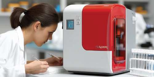
At our Vet Clinic in Armidale, one of the fastest and most effective ways to get those facts is through a process called Cytology. It allows us to look at the problem on a microscopic level, often giving us an answer before you even leave the consult room.
Here is what this test is, how we do it, and why it is so valuable.
What is Cytology?
Simply put, Cytology is the study of loose cells.
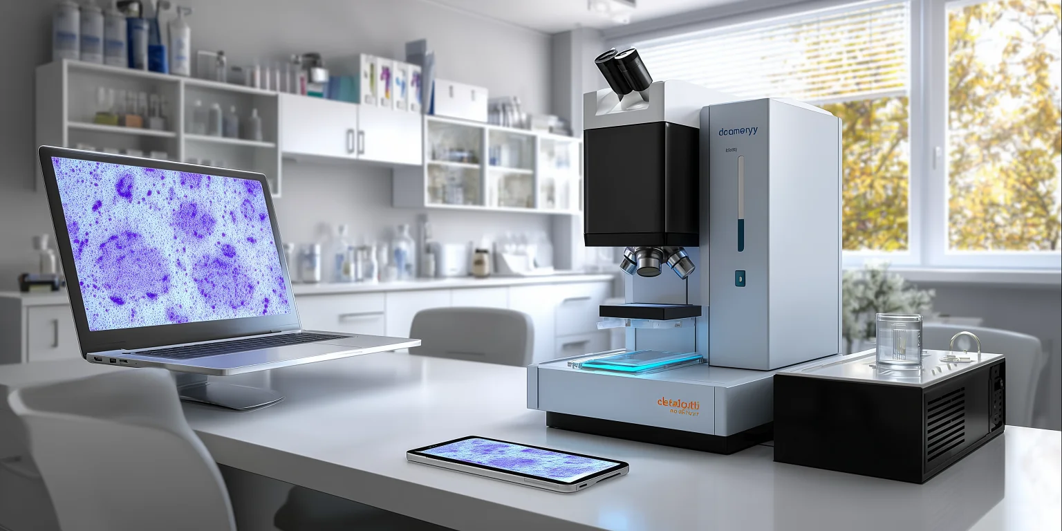
Unlike a biopsy (which involves cutting out a chunk of tissue to look at the “architecture”), cytology involves taking a tiny sample of cells and spreading them onto a glass slide. We then stain them with special dyes and examine them under a high-powered microscope.
It helps us answer three main questions:
Is it Inflammation? (An allergic reaction or immune response).
Is it Infection? (Bacteria, yeast, or fungi).
Is it Neoplasia? (A benign growth or a malignant cancer).
It’s Not Just for Lumps
While we often use cytology for lumps, it is actually a versatile tool for many issues:
The “Lump Check” (Fine Needle Aspirate): We insert a small needle into a mass to suck out some cells. This is great for distinguishing a harmless fatty lump (Lipoma) from something nastier like a Mast Cell Tumor.
Ear Infections: If your dog has itchy ears, we take a swab of the wax. Cytology tells us if the infection is Yeast or Bacteria (and what shape the bacteria are). This is crucial because the medication for yeast does not work on bacteria, and vice versa!
Skin Issues: We might use clear sticky tape to lift cells off a hot spot to check for mites or surface infections.
What to Expect During Your Visit
One of the best things about cytology is that it is minimally invasive.
1. The Collection
For Lumps: We perform a Fine Needle Aspirate (FNA). It feels very similar to receiving a vaccination. Most pets don’t even flinch, and sedation is rarely required.
For Ears/Skin: We use cotton swabs or medical tape. This is completely painless, though some dogs find the ear tickle a bit annoying!
2. The “Squash” & Stain
Once we have the sample, we spray it onto a glass slide. If it is an FNA, we use a technique to “squash” the cells into a single layer so light can pass through them. We then dip the slide into three different staining liquids (turning the cells pink and purple/blue).
3. The Microscope
In-House: For ear infections and simple lumps, our vets examine the slides right here in the clinic. We can often give you an answer in 10–15 minutes.
Pathologist Review: For complex or ambiguous lumps, we send the slides to a specialist veterinary pathologist. They have trained for years to identify rare cell types. These results usually take 1–3 days.
If you have found a lump on your pet, or if they are shaking their head with ear trouble, book an appointment at North Hill Veterinary Clinic today. Let’s take a closer look.